When it comes to vascular diagnostics, LSB-RVT testing plays a key part. It gives clear insight into venous health. Closer word links make each idea easy to grasp, so we use simple, tight connections.
In this article, we explain what LSB-RVT testing is, why it matters, how it is done, and how to read its results. This guide helps both clinicians and patients. Whether you are a doctor needing a review or a patient seeking answers, these points guide you with care.
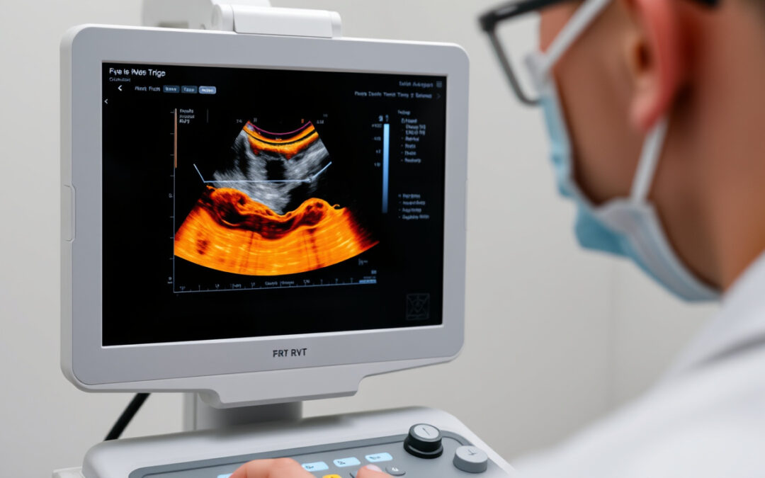
What Is LSB-RVT Testing?
LSB-RVT testing means Lower Segmental Branch–Right Venous Thrombosis testing. The test views the lower segmental branch veins in the leg. It finds blood clots (thrombi) that may hide in these small branches. Its focus is on places where a standard Doppler ultrasound may not see enough. Using close word ties helps us see the test’s role clearly in finding deep vein thrombosis (DVT).
This test is important because it checks for clots in small vein branches. Clots may lead to serious problems like pulmonary embolism if they go unnoticed.
Why Is LSB-RVT Testing Important?
Veins in your legs can get blocked and cause big health issues. Standard venous scans give a good overview but sometimes miss small clots. Here, LSB-RVT testing helps with these tasks:
- It detects clots early in small vein branches.
- It clears up unclear ultrasound findings.
- It watches patients with high clot risk.
- It guides better blood thinner treatment.
How Does LSB-RVT Testing Work?
LSB-RVT testing uses modern ultrasound and clear imaging steps. The method shows veins that are hard to see in routine scans. We connect each word closely to make steps simple:
-
Patient Preparation:
The patient wears loose clothes. They may remove items that cover the legs. No fasting or extra steps are needed. -
Positioning:
The patient lies on their back or in a semi-reclined position. The leg is bent or turned slightly outward to show the veins clearly. -
Ultrasound Imaging:
A high-frequency linear probe is used. The technician scans the lower limb veins one by one. Special color Doppler imaging shows normal flow or blockages. -
Compression Testing:
The probe applies light pressure. The test checks if a vein can squeeze normally. Poor squeeze means a clot may be present. -
Doppler Flow Analysis:
Blood flow patterns are checked. This step shows any blockages in the veins.
The test usually takes 30 to 45 minutes. It is safe, pain-free, and does not use radiation.

Who Should Undergo LSB-RVT Testing?
Doctors recommend LSB-RVT testing for those with signs of venous problems. Look out for these symptoms:
- Swelling, pain, or tenderness in the lower leg.
- Skin that looks discolored or feels warm.
- A history of deep vein thrombosis or pulmonary embolism.
- Recent surgery or injury with long rest.
- Coagulation issues or cancer.
- Unexplained leg swelling or suspected venous weakness.
Physicians may also use this test to check treatment progress or assess clot risk before procedures.
Interpreting LSB-RVT Test Results
Doctors review the test by looking at vein squeeze, flow patterns, and any clots. The answers come in a clear form:
- Normal findings:
Veins compress well, blood flows normally, and no clot is seen. - Partial thrombosis:
The vein does not compress fully, showing a partial blockage. - Complete thrombosis:
The vein does not compress. There is no blood flow, and a clot is visible.
Early detection by LSB-RVT testing leads to quick treatment with blood thinners, lowering the risk of serious problems.
Advantages of LSB-RVT Testing
- High Sensitivity:
It finds clots in small, hard-to-see vein branches. - Non-Invasive:
No pain, no needles, and no radiation. - Rapid Results:
Results show immediately to guide quick decisions. - Repeatable:
The test can be done many times if needed.
Limitations and Considerations
LSB-RVT testing is very useful. Yet, it has limits:
- Operator Dependence:
The test works best when done by an expert. - Limited Availability:
Not every center has the right machine or trained staff. - Anatomical Complexity:
Different body anatomies may make scanning harder.
Doctors must consider the full patient picture. They may use CT venography or MRI to double-check results.
Tips to Prepare for Your LSB-RVT Test
Patients can make the test go smoothly by following these tips:
- Wear loose, comfortable clothes to expose the legs.
- Tell your doctor about all medical conditions.
- Arrive on time to avoid delays.
- Drink plenty of water unless told otherwise.
Frequently Asked Questions (FAQs) about LSB-RVT Testing
Q1: What exactly does LSB-RVT testing detect?
It finds blood clots in the lower vein branches, often in the right leg. The test helps diagnose deep vein thrombosis where other exams might not see it.
Q2: Is LSB-RVT testing painful or risky?
No, it is a safe and non-invasive ultrasound test. It usually causes no pain and carries little risk, with no radiation.
Q3: How soon can I expect results after LSB-RVT testing?
Results are shared immediately. Your doctor can review them during the test visit for quick action.
Conclusion
Understanding LSB-RVT testing gives clear insight into finding and managing venous clots. The test is precise, non-invasive, and fast. It helps sort out unclear symptoms and guides timely treatment. This method can protect patients from serious risks linked to thrombosis.
If you or someone you care about shows signs of venous disorders, talk to your healthcare provider about LSB-RVT testing. It might be the first step toward early diagnosis and effective treatment.
For more in-depth clinical guidelines on testing for vein clots, please refer to resources from the American College of Radiology (source).

